|
||||||||||||||||||||||||||||||||||||||
|
OMA International Group Mobile: 00 964 77 0 444 6566 |
||||||||||||||||||||||||||||||||||||||
|
||||||||||||||||||||||||||||||||||||||
| شركة أوما الدولية | ||||||||||||||||||||||||||||||||||||||
| OMA International Trading | ||||||||||||||||||||||||||||||||||||||
 |
||||||||||||||||||||||||||||||||||||||
|
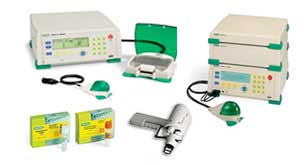
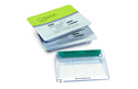
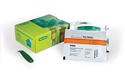
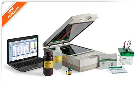
Specifications
Biologics
Analysis Workflow™ |
||||||||||||||||||||||||||||||||||||||
 |
||||||||||||||||||||||||||||||||||||||
|
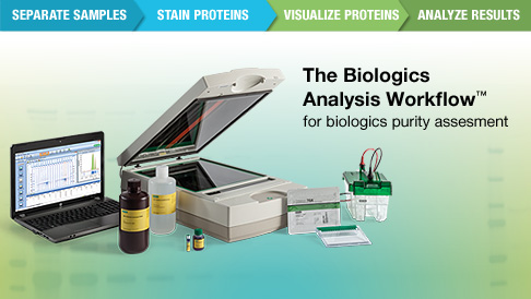
The Biologics Analysis Workflow is a suite of products designed and
validated to assess the purity or identity of biological products in a cGMP
regulatory environment. The Biologics Analysis Workflow provides:
Increased throughput
Simplified, sensitive gel staining
Accurate and reproducible imaging
Easy data analysis
The Biologics Analysis Workflow includes a set of integrated
protein electrophoresis solutions for each step of the workflow:
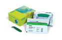
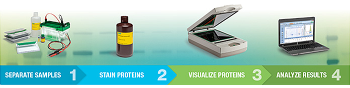
Step 1: Separate samples
Criterion™ TGX™ precast gels — increased throughput with fast run
times and more sample loading wells
The unique properties of TGX chemistry reduce run times to as
little as 20 minutes. These gels have a 1-year shelf life and utilize the
standard Laemmli buffer system. The midi-sized Criterion format allows you to
load up to 26 samples while maintaining the required resolution. Additionally,
the characteristic upper buffer chamber reduces the amount of running buffer
required.
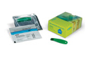
Step 2: Stain proteins
QC Colloidal Coomassie stain — highly sensitive, reliable,
end-point stain designed for quantitative analysis of proteins
QC Colloidal Coomassie stain enables the detection of nanogram
levels of protein. The colloidal chemistry improves the protein-to-dye binding
ratio, reducing background staining and improving sensitivity. QC Colloidal
Coomassie stain is an end-point stain that was designed
and
optimized for quantitative analysis with the GS-900™ densitometer. The stain is
ready to use without mixing or the addition of other reagents. The stain is
formulated with ethanol instead of methanol, eliminating the need for hazardous
waste disposal.
Step 3: Visualize proteins
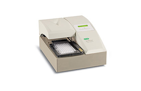
GS-900™ calibrated densitometer —
reproducible imaging of protein gels, blots, and film across a wide dynamic
range
To ensure the accuracy of each scan,
the GS-900 Calibrated densitometer contains internal optical density tablets
that are scanned and used for calibration before each run. The calibration of
the GS-900 can be validated with the GS-900 calibrated densitometer
installation and qualification kit, which utilizes an external target to
confirm accuracy of the internal target, ensuring the accuracy and
reproducibility of the GS-900 densitometer. With red/green/blue tricolor CCD
technology, the GS-900 calibrated densitometer enables the imaging and
quantitation of film, blots, and gels stained with a wide variety of stains.
The wide dynamic range permits the quantitation of proteins from 0–3.4 OD.
Step 4: Analyze results |
||||||||||||||||||||||||||||||||||||||
 |
||||||||||||||||||||||||||||||||||||||
|
Image Lab™ image analysis software —
effortless analysis of samples with an intuitive, user-friendly interface
Gel analysis is greatly simplified
with Image Lab software. The ability to automate analyses saves time and
ensures consistent analysis from run to run. Image Lab software includes
detailed tutorials and requires no previous imaging experience to produce
optimum gel and blot images. The flexible system allows users to easily modify
analysis settings to match SOPs and fine-tune band detection, background level,
and other settings. Image Lab software makes reporting easy with the abil |
||||||||||||||||||||||||||||||||||||||
 |
||||||||||||||||||||||||||||||||||||||
|
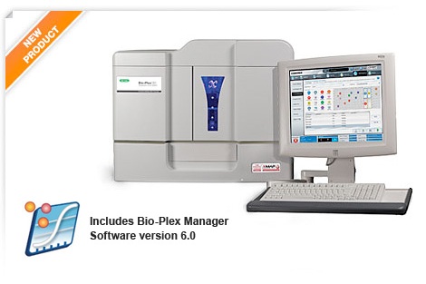
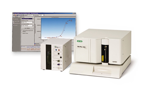
Bio-Plex®
3D suspension array system Bio-Plex instruments include the
Bio-Plex MAGPIX, Bio-Plex 200, and Bio-Plex 3D systems, Bio-Plex Pro™ wash
stations, as well as maintenance, calibration, verification, and validation
kits. |
||||||||||||||||||||||||||||||||||||||
|
||||||||||||||||||||||||||||||||||||||
|
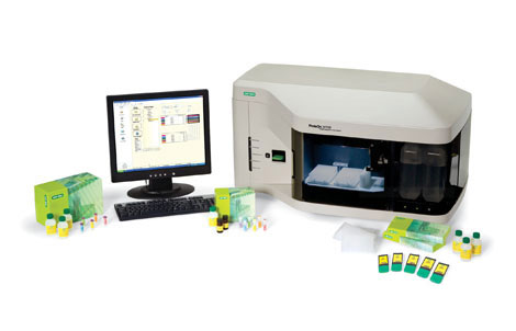
1- The Bio-Plex® 3D
suspension array system is the next generation multiplexing platform based on
xMAP technology. Expanded multiplexing capability, faster time to results, and
automation capability make it the platform of choice for high-throughput
testing for nucleic acid and protein applications.
·
Measure up to 500 unique analytes
in a single sample
·
96- and 384-well plate capability
·
Plate read times twice as fast as
the Bio-Plex/Luminex 200 system
·
Robotics interfacing capabilities
·
LIS-compatible software
·
Compatible with the magnetic and
nonmagnetic assays
·
Bio-Plex Manager™ software
version 6.0 for data analysis
·
Onsite training Bio-Rad's Bio-Plex assays can be
used with this system. Please check the Bio-Plex 3D Assay Compatibility Chart
in the overview before buying assays of interest. Bio-Plex 3D Instrument Optional Upgrades for the Bio-Plex 3D system 21CFR Part 11 Module Specifications
|
||||||||||||||||||||||||||||||||||||||
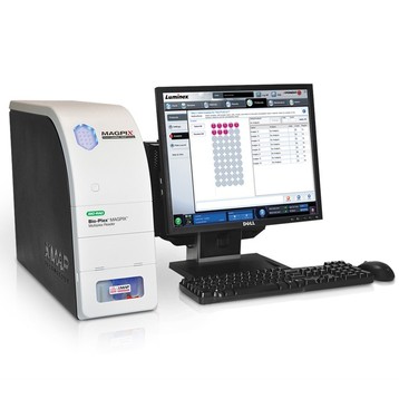
| Bio-Plex
MAGPIX |
||||||||||||||||||||||||||||||||||||||
|
||||||||||||||||||||||||||||||||||||||
|
The Bio-Plex MAGPIX multiplex reader is a compact, robust system
for magnetic bead–based immunoassays. This multiplex reader is capable of
reading assays designed on magnetic xMAP (MagPlex) beads, compatible with
Bio-Plex Pro™ magnetic assays.
Key Features of the Bio-Plex MAGPIX Multiplex Reader
Get more data by reading up to 50 analytes per sample
Simple and convenient ELISA-like workflow
Benefit from sample and cost savings with Bio-Plex Pro magnetic
bead–based assays in single or multiplex format
Provide improved multiplex productivity and convenience with
magnetic bead–based assays
Save bench space with a compact footprint
Conserve precious resources with an affordable low maintenance
system
Obtain the same quality results achieved on the Bio-Plex®
200 or Bio-Plex 3D systems
Take advantage of automated instrument management and other
software features:
Automatic loading of recommended start-of-day maintenance routines
based on current instrument status
Performance monitoring during data acquisition alerts the user to
performance issues and loads recommended maintenance to resolve issues
Simple step-wise troubleshooting
Benefits of Bio-Plex Manager MP Software
Detection and resolution of performance issues ensures data quality
Confidence in proper instrument maintenance leads to confidence in
results
Highest quality results maximize the value of your experiments
Simple integration with
Bio-Plex
analysis software to quickly assemble and understand your data
Simple interface and built-in guidance enable a user of any
experience level to easily run the Bio-Plex MAGPIX multiplex reader
Components of the Bio-Plex MAGPIX Multiplex Reader
Bio-Plex MAGPIX instrument
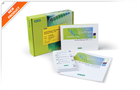
Bio-Plex Manager MP software
Bio-Plex Manager software
PC with xPONENT 4.2 acquisition software installed
Probe height adjustment plate
Calibration kit
Verification kit
2 drive fluid cartridges
2 waste containers
Getting started guide
Upgrade quick guide
Related Products
Bio-Plex Data Pro™ software for data management and visualization
Bio-Plex Manager™ MP software is included with Bio-Rad’s Bio-Plex MAGPIX
reader
More Information
Visit the
Bio-Plex software resource page
for the latest
software updates, information, and standard lot values. Specifications
|
||||||||||||||||||||||||||||||||||||||
|
Bio-Plex Pro™ Wash Stations |
||||||||||||||||||||||||||||||||||||||
|
||||||||||||||||||||||||||||||||||||||
|
Bio-Plex Pro wash stations eliminate manual wash steps from Bio-Plex
assays, making Bio-Plex assays as easy as ELISAs. The wash stations are
specifically designed to perform Bio-Plex assay wash steps, but are compatible
with any standard xMAP assay.
Benefits of Bio-Plex Pro wash stations include:
Improved lab productivity
Reliable and reproducible results
Optimized on-board protocols
Two versions of the wash station are available:
Bio-Plex Pro wash station — incorporates
a magnetic plate carrier for reliable, hands-free wash steps on magnetic
bead-based assays (Bio-Plex Pro and MagPlex assays)
Bio-Plex Pro II wash station — includes both
a magnetic carrier and an interchangeable vacuum manifold for hands-free wash
steps on any xMAP assay (magnetic and nonmagnetic bead-based assays)
Both options include preset wash programs that have been optimized
for Bio-Plex assays. Using Bio-Plex Pro wash stations in an assay workflow can
reduce manual intervention and help decrease variability between independent
experiments. Specifications
Protein interaction analysis
|
||||||||||||||||||||||||||||||||||||||
|
||||||||||||||||||||||||||||||||||||||
|
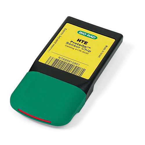
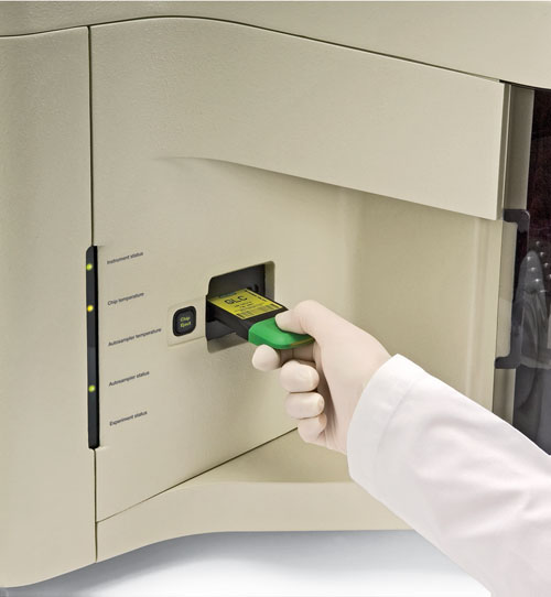
The ProteOn™ XPR36 protein interaction array system is an SPR
optical biosensor that provides real-time data on the affinity, specificity, and
interaction kinetics of protein interactions. Using XPR technology*, a unique
approach to multiplexing, this system generates a 6 x 6 interaction array for
the simultaneous analysis of up to six ligands with up to six analytes. The
ProteOn XPR36 system increases the throughput, flexibility, and versatility of
experiment design, enabling the completion of more experiments in less time: Analyze up to 36 different protein interactions in a single run, on
a single chip Perform a complete kinetic analysis in a single run Measure a variety of experimental conditions simultaneously in
parallel Screen multiple panels of analytes See the
ProteOn Technology and Applications videos for more
information about how the ProteOn system works and how it can be applied to drug
discovery and to address important experimental questions. The ProteOn XPR36 protein interaction analysis system includes all
of the components required for interaction analysis: instrumentation,
ProteOn Manager™ software,
ProteOn sensor chips,
buffer and reagent kits, and
protocol development kits. User-friendly ProteOn Manager software uses a flexible, guided
approach to coordinate instrument control, experiment setup, and data analysis. Key Features and Benefits Novel 6 x 6 array design Real-time data acquisition Analysis of interaction kinetics, binding affinity, and analyte
concentration No radiochemical or fluorescent labels needed Integrated system Applications and Uses Antibody characterization and development Large and small molecule drug development Protein interface analysis Protein complex and cascade analysis The ProteOn XPR36 protein interaction array system is covered by
Bio-Rad patents, including United States patent numbers 8,111,400, 8,105,845,
7,999,942, and 7,443,507. This product or portions thereof is manufactured and sold under
license from GE Healthcare under United States patent numbers 5,492,840,
5,554,541, 5,965,456, 7,736,587, and 8,021,626, and any international patents
and patent applications claiming priority. Specifications
|
||||||||||||||||||||||||||||||||||||||
|
||||||||||||||||||||||||||||||||||||||
|
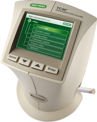
TC20 Automated Cell counter |
||||||||||||||||||||||||||||||||||||||
|
||||||||||||||||||||||||||||||||||||||
|
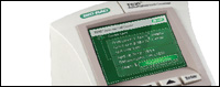
The TC20 automated cell counter counts mammalian cells in one simple
step using its innovative auto-focus technology and sophisticated cell counting
algorithm to produce
accurate cell counts in less than 30 seconds.
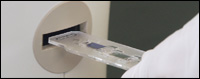
Upon insertion of a counting slide, the TC20 cell counter rapidly provides a
total cell count (with or without trypan blue staining) and
assesses cell viability via trypan blue exclusion. Key Features and Benefits Compatible with a broad range of
cell sizes and types — counts cell lines, primary cells
(from tissue or blood), and stem cells Innovative auto-focus technology — removes the variation associated with
manual focusing and leads to precise cell counts in 30 seconds Cell size gates — user selects a population of interest in
complex samples, such as primary cells, or lets the cell counting algorithm do
all the work Cell viability — analyzes cells accurately using
multifocal plane analysis Easy to archive and analyze — stores up to 100 counts in the onboard
memory for access any time, or use the optional
TC20 data analyzer software on your PC to further analyze exported cell
images Getting all the data you need about
your cell cultures is fast and easy; the TC20 cell counter and disposable
counting slides eliminate the need for tedious setup, cleaning, or maintenance.
The TC20 cell counter is simple and intuitive to use
Check out the TC20 automated cell counter
introduction video.
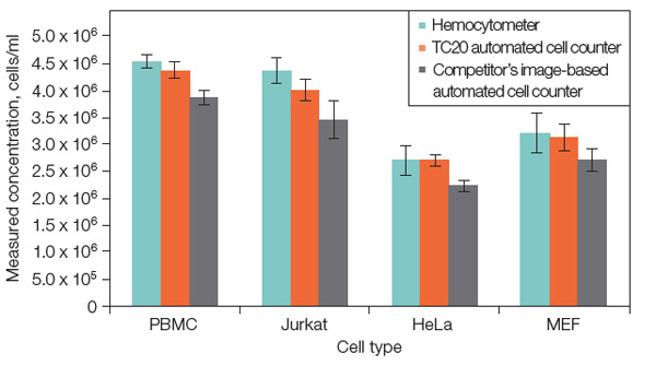
Get Accurate and Reproducible Results The TC20 automated cell counter uses microscopy with auto-focus that
analyzes multiple focal planes to identify the best plane. Without requiring any
user input, the sophisticated cell counting algorithm uses the image acquired
from the best focal plane to identify cells and exclude debris, thereby
calculating the total cell count. The auto-focus leads to highly reproducible cell counts with reduced
user-to-user variability compared to a hemocytometer and cell counters with
manual focus. Using auto-focus instead of subjective manual focusing is
especially important when assessing cell viability because an incorrectly
selected focal plane will lead to inaccurate results. Unlike cell counters based on electrical impedance (Coulter
principle), the TC20 image-based analysis allows the user to view the cells in
real time, providing visual proof that the TC20 cell counter has correctly
identified cells. The TC20 automated cell counter provides accuracy comparable to
results obtained with a hemocytometer. It can count cells with a 6–50 μm cell
diameter and within a broad concentration range of 5 x 10⁴–1 x 10⁷
cells/ml, which eliminates the need to dilute cells, thus reducing errors
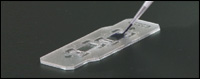
associated with sample dilutions prior to counting. The counter uses disposable counting slides with a patent-pending
design that ensures even distribution of cells throughout the counting chamber,
regardless of the user's pipetting style, leading to accurate and consistent
cell counts. For complex samples composed of multiple cell populations, such as
primary cells, users can adjust the cell size gates to define the population of
interest that will be counted. When counting multiple sample replicates, the
TC20 cell counter can save the position of the cell size gates and apply them to
subsequent counts. |
||||||||||||||||||||||||||||||||||||||
Cell Viability Minimize Your Sample Preparation The broad concentration range of the TC20 cell counter eliminates
the need to dilute cells prior to counting, reducing the errors associated with
sample dilutions that may be necessary when counting cells by other methods. The
counting algorithm discriminates and counts individual cells within clusters of
up to five cells, providing accurate counts without the need to extensively
declump cells prior to loading.
|
||||||||||||||||||||||||||||||||||||||
|
||||||||||||||||||||||||||||||||||||||
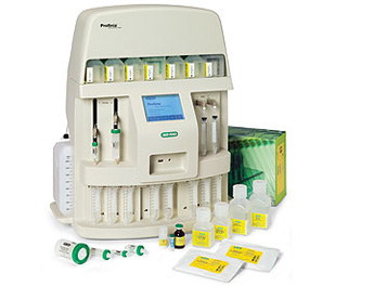
| Profinia™ Affinity Chromatography Protein Purification System |
||||||||||||||||||||||||||||||||||||||
|
||||||||||||||||||||||||||||||||||||||
|
The Profinia protein purification
instrument is compact and easy to use. Each compartment of the instrument is
clearly labeled for fast setup. The buffer compartment allows simple
installation of buffers — numbered positions on the instrument match run
diagrams and prepackaged reagent labels. The sample compartment is capable of
running two samples in series. Both compartments have sipper tubes for the
uptake of buffers and samples. The cartridge compartment offers two-step
purification capability and uses luer fittings to simplify cartridge connection.
Waste collection and diluent bottles are in view and accessible on the sides of
the instrument. The fraction collection and sample compartments allow quick
installation of standard collection tubes. A central compartment provides
convenient storage for a stylus and access to a USB flash drive, which allows
you to export run data.
The instrument has a large
touch-screen user interface, where you can select options — by finger or stylus
— to navigate from screen to screen. Intuitive, user-friendly help functions
guide you through the setup and post-run functions of preprogrammed optimized
purification methods. The methods and buffer sets include cleaning protocols
that maintain the system and prepare the cartridges for future use, ensuring
that the Profinia system stays in optimal running condition. This combination of
features allows you to set up quickly, walk away, and come back to results in as
little as 30 min.
·
Optimized methods, cartridges, and
buffer kits eliminate time spent on method development, troubleshooting, and
reagent preparation
·
Built-in reproducibility is
achieved with automated system pumps, UV and conductivity detectors, and
programmed cleaning methods
·
Histidine (His)-tagged,
GST-tagged, affinity, and desalting/buffer exchange methods deliver purity and
yield comparable to other techniques, and require only a fraction of the time to
obtain protein ready for experiments
·
Automatic UV peak detection
diverts eluted target protein from cartridges to a fraction collection tube for
unattended operation
·
Easy-access fraction collection
and sample compartments hold 15 or 50 ml conical collection tubes
·
Benchtop or coldroom operation
allows you to control purification conditions; optional cooling accessory keeps
samples and fractions cold for benchtop operation
·
Additional capabilities let you
work with a pH monitor that runs in real time with Profinia software
·
Specifications for Profinia™
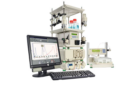
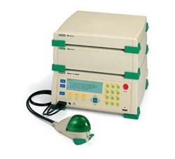
BioLogic DuoFlow 10 System |
||||||||||||||||||||||||||||||||||||||
|
||||||||||||||||||||||||||||||||||||||
|
The BioLogic DuoFlow™ family of chromatography systems offers
flexibility with multiple system configurations, many optional upgrades, and a
common software platform that is intuitive and easy to follow. These systems can
be used on the laboratory bench or in a coldroom. The BioLogic DuoFlow 10 system includes an F10 workstation for a
flow rate of 0.01–10 ml/min* at 3,500 psi (233 bar, 23 MPa). A Model MX-1 mixer,
3-tray rack, AVR7-3 sample inject valve, fittings kit, UV detector with 5 mm
flow cell and 254/280 nm filters, conductivity monitor, starter kit, UNO®
Q1 column, and instructions are also included. A Dell PC controller enables easy communication with the workstation
and peripheral devices via an external USB Bitbus communicator. The controller
includes the Windows XP operating system, application software, keyboard, mouse,
and high-resolution flat-panel monitor. BioLogic DuoFlow modular components allow the system to meet both
laboratory space and application requirements; as requirements change, systems
may be easily reconfigured and seamlessly upgraded with increased functionality,
such as higher flow rates, sophisticated detection capabilities, pH monitoring,
column scouting, and buffer blending. * To double the flow rate to up to 20 ml/min, use the BioLogic
DuoFlow Maximizer™ valve system. The optional F40 pump kit expands pumping
capabilities to 40 ml/min Specifications
|
||||||||||||||||||||||||||||||||||||||
|
||||||||||||||||||||||||||||||||||||||
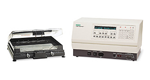
|
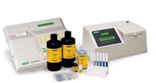
The SmartSpec Plus spectrophotometer has a wider range of features
and functions than many other benchtop spectrophotometers, offering performance,
stability, and functionality at an affordable price. The UV/visible SmartSpec Plus spectrophotomer has a working
wavelength range of 200–800 nm. It is the perfect tool for routine applications
such as: Quantitation of DNA, RNA, and oligonucleotides Quantitation of proteins via the Bradford, Lowry, and BCA assay
methods Monitoring bacterial culture growth Simple kinetic assays Wavelength scans with peak detection A simple, menu-driven interface simplifies assays and provides
answers to common sample computations at the touch of a button. Conversion
factors can be stored and modified. The SmartSpec Plus spectrophotometer is
capable of performing calculations and providing results such as: A260/A280 ratio for nucleic acid purity Quantitation that takes dilution factors into account Sample concentration in µg/ml (additionally in pmol/µl for
oligonucleotides) Molar extinction coefficient and molecular weight of
oligonucleotides At the end of an assay, a report may be printed that shows the
user's identification, date, and results. Nucleic Acid Quantitation The SmartSpec Plus spectrophotometer also simplifies the
quantitation of DNA and RNA oligonucleotides. When you enter the sequence,
length, or composition, the spectrophotometer will display sample concentration
in µg/ml and pmol/µl and automatically perform calculations of molar extinction
coefficients and molecular weights. Protein Quantitation Standards can be analyzed in groups of up to 9 replicates Up to 10 standard curves can be stored under user-assigned names Mean and standard deviation values are automatically calculated for
each replicate group Printable report includes a standard curve with r2 value Additional Features Built-in printer Xenon flash lamp extends lamp life and reduces maintenance costs User interface with choice of 6 languages: English, French, German,
Italian, Japanese, or Spanish Spectral scans from 200 to 800 nm Easy-to-use, menu-driven operation Compact, space-saving design Specifications |
||||||||||||||||||||||||||||||||||||||
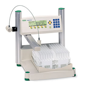
Transfection A broad suite of transfection tools
is available for nucleic acid delivery into a wide range of cell types.
Instruments and kits are designed for a variety of applications, such as RNAi
gene knockdown and primary cell analysis. |
||||||||||||||||||||||||||||||||||||||
|
||||||||||||||||||||||||||||||||||||||
|
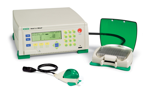
The Gene Pulser MXcell plate-based
electroporation system delivers molecules
efficiently into mammalian cells — especially
into primary and difficult-to-transfect cells.
The Gene Pulser MXcell system can be used to
electroporate as few as 105 cells by using a
96-well plate or as many as 106 to 107 cells in
a 12-well plate or in a cuvette. The system's
enhanced user interface contains preset
protocols to easily adapt to new or existing
transfection conditions. |
||||||||||||||||||||||||||||||||||||||
 |
||||||||||||||||||||||||||||||||||||||
|
Key Benefits and Features
Delivery of any molecule into primary
and other mammalian cells — transfect siRNA, DNA, and other molecules in a
completely open format
Preprogrammed protocols — designed
for rapid optimization to increase transfection efficiency and viability of any
mammalian cell type, including primary and difficult-to-transfect cells
Fully programmable — modify protocols
for your specific needs to obtain better gene delivery
Choice of electroporation plate or
cuvette — electroporate a limited number of primary cells or a larger number
when scaling up
Fast pulse time — minimize cell
handling
Protocols for primary and mammalian
cells — browse Bio-Rad's expanding Transfection Protocol Library, which includes
electroprotocols describing recommended starting conditions for most mammalian
cells
Compatible with any electroporation
buffer — use with Gene Pulser® electroporation buffer for efficient gene
delivery while maintaining cell viability in mammalian cells
Built-in safety features — arc
protection for detecting precise location of well arcing and resistor pulse
modulation for controlling parallel resistance. |
||||||||||||||||||||||||||||||||||||||
|
||||||||||||||||||||||||||||||||||||||
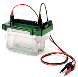
|
Mini-PROTEAN® Tetra Cell - Electrophoresis |
||||||||||||||||||||||||||||||||||||||
|
||||||||||||||||||||||||||||||||||||||
|
The versatile, easy-to-use Mini-PROTEAN® Tetra cell is ideal for
vertical mini gel electrophoresis. This electrophoresis cell accommodates one to
four Mini-PROTEAN precast or handcast gels, providing flexibility for your
research needs. The Mini-PROTEAN Tetra cell eliminates tedious assembly
procedures and has a patented sealing mechanism* that prevents assembly errors
during handcasting. Features and Benefits Run one to four precast or handcast mini gels in less than an hour Can also be used with the Mini Trans-Blot® electrophoresis cell for
western blotting Thermoplastic casting gaskets form a tight seal with the glass
plates to ensure leak-free casting Casting frames** with simple cam closure provide precision alignment
on any flat surface Side-by-side casting stand** allows access to both gels
simultaneously, and a spring-loaded lever creates a tight seal against the
thermoplastic rubber gasket Run 2-D gels in less than a day Applications and Uses Polyacrylamide gel electrophoresis (PAGE or SDS-PAGE) 2-D gel electrophoresis Screen new samples Evaluate sample preparation conditions
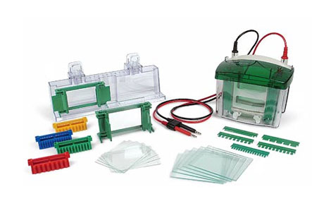
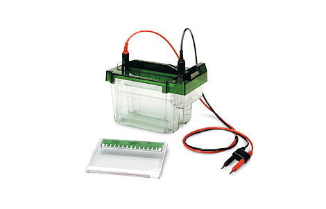
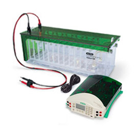
Criterion™ Cell and Power Supply |
||||||||||||||||||||||||||||||||||||||
|
||||||||||||||||||||||||||||||||||||||
|
The Criterion™ cell is a midi
electrophoresis cell dedicated to running one or two Criterion gels, which are
wider and longer than traditional mini gels. With a single Criterion gel, you
can run up to 26 samples in less than 1 hr or accommodate 11 cm ReadyStrip™ IPG
strips for 2-D applications. The Criterion cell accommodates Criterion precast
or handcast gels. Features and Benefits
·
Compact size requires only 1 L
running buffer
·
Integrated cassette buffer chamber
simplifies assembly and ensures that the system will never leak
·
Safe, easy-to-open cassettes use a
built-in wedge on the lid to open gel cassettes in a single step
·
Locator slots built into the tank
walls allow users to easily and quickly slide cassettes into position
·
Ability to separate more samples
in fewer runs and resolve more proteins
·
Run 2-D gels in less than a day Applications and Uses
·
Polyacrylamide gel electrophoresis
(PAGE or SDS-PAGE)
·
2-D gel electrophoresis
·
Screen new samples
·
Evaluate sample preparation
conditions
Pulsed Field Gel Electrophoresis Systems |
||||||||||||||||||||||||||||||||||||||
|
||||||||||||||||||||||||||||||||||||||
|
The CHEF Mapper®
XA system incorporates patented FIGE and AFIGE* technologies for superior
resolution in the range of 100 bp to 10 Mb, in addition to the features of the
CHEF-DR® II and the CHEF-DR III systems. The CHEF Mapper XA system also includes
algorithms for deriving separation conditions. This system is ideal for both the
PFGE novice and the seasoned expert.
Each CHEF Mapper
XA system is supplied with a power module, embedded auto-algorithm for protocol
optimization, interactive algorithm program disk, electrophoresis cell, cooling
module, variable-speed pump, and accessory kit.
Automation
The CHEF Mapper
XA system offers two ways to optimize your separations. The unique
auto-algorithm can automatically select optimal separation conditions,
integrating 11 key variables and implementing starting separation conditions.
Protocols can be refined using the Windows operating system-based interactive
algorithm, which lets you specify several run variables simultaneously to derive
optimal separation protocols.
Customization
Busy
laboratories need equipment that can store and readily access key separation
conditions. The CHEF Mapper XA system can store up to 99 programs.
Application Versatility
The CHEF Mapper
XA system is the most flexible of the PFGE units. The CHEF Mapper XA system
achieves higher resolution with greater speed and accuracy than any other PFGE
system, making it ideal for all PFGE applications. The CHEF Mapper XA system
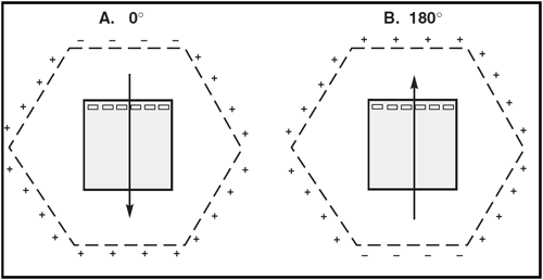
lets you choose any pulse angle from 0 to 360°, which allows optimal separation
of both chromosomal and plasmid DNA with one system. Accurate sizing of
fragments requires an expanded linear range of separation. Switch-time ramps
increase the mobility of fragments by gradually changing the switch times during
the course of a run. Nonlinear (for example, concave or convex) ramps change the
switch-time increments from the start to the end of a run.
These nonlinear
ramps separate fragments linearly from 50 to 700 kb, yielding more precise
fragment size measurements. The multistate mode of the CHEF Mapper XA system
enhances resolution in selected fragment size ranges. Each vector (angle of
pulse) can be assigned its own voltage (field intensity) and its own switch time
(duration of pulse). Up to eight different states can be combined into one run
to optimize the separation of subsets of fragments in the sample. The
application of secondary pulses of defined voltage, duration, angle, and
frequency can enhance the separation and resolution of very large DNA molecules
by releasing DNA caught in the gel matrix. |
||||||||||||||||||||||||||||||||||||||
 |
||||||||||||||||||||||||||||||||||||||
 |
||||||||||||||||||||||||||||||||||||||
| .......................................................................................................... | ||||||||||||||||||||||||||||||||||||||
|
Category Products
Category Products
|
||||||||||||||||||||||||||||||||||||||
| Download | ||||||||||||||||||||||||||||||||||||||
|
|
||||||||||||||||||||||||||||||||||||||
|
|
||||||||||||||||||||||||||||||||||||||
| OMA International Group | ||||||||||||||||||||||||||||||||||||||
|
For more information, please visit our website (www.omatrade.com).
Contact Information: Mobile: 00 964 77 0 444 6566 Email: sales@omatrade.com , support@omatrade.com, zaid@omatrade.com |
||||||||||||||||||||||||||||||||||||||
| Al-Shareif Building, Street 9, District 102, Alweyah Baghdad-Iraq - P.O BOX: 3056 | ||||||||||||||||||||||||||||||||||||||
|